Dan Milkie
I am a Senior Scientist in Nobel Laureate Eric Betzig’s group at the Janelia Research Campus. My interests are in tool building for science, including super resolution light microscopy and adaptive optics.
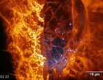
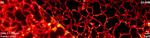
Cells did not evolve to live on the glass coverslip nor under the burning intensity of high power lasers. Unlocking what cells and biology are doing dynamically and in their native environment is key to understanding how complex biologic systems work.
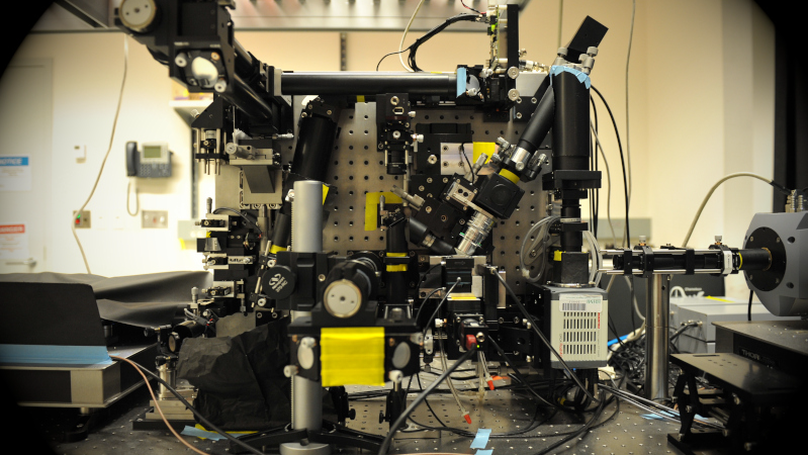
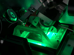
I create the microscope hardware and software control systems for the Betzig Group and others, and bring to bear any new technology we can leverage (FPGA, Spatial Light Modulators, Deformable Mirrors, Piezo stage, >GB/s cameras,… ).
I build, first, for the hands of power users. Next comes a large investment: taking their achieved wisdom and baking it into new hardware and software versions, but it’s totally worth it. The pay-off comes with wide impact distribution, commercialization, and use by non-specialists. Tech can’t be a limited to a unicorn machine in a single lab doing a hero experiment.
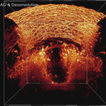
Volumetric imaging microscopy platforms deliver a firehose of 4D data, which generate a host of new, enticing challenges (data transfer, storage, curation, processing, visualization, ML analysis) which are the catnip to my future.
Download my resumé.
View my publications.
- Microscopy
- Machine Vision
- LabVIEW
- Python
- Automation
- Efficiency
PhD in Physics, 2008
University of Pennsylvania
BSc in Physics, 2002
The College of William and Mary
Experience
Created the hardware control system and control software for all Betzig microscopes and few others
- Lattice Light Sheet
- AO Lattice Light Sheet
- AO Bessel
- Structured Illumination Micrsocope
- Opposing Objective Microscope
- Cryo-SIM/-PALM
- MOSAIC (Adaptive Optics added to everything we can in one scope)
Grew a small machine vision-centric company, while solving industrial part-inspection tasks and scientific research automation challenges.
- Built systems to inspect: Dinnerware, truck brakes, agriculture seeds, optical fiber, mass flow controllers,…
- Point-cloud 3D laser height mapping, bright & dark field imaging, Area scan cameras, and Conveyor-based line-scan camera systems
- Oversaw projects, contracts, budgets, sales, small teams, recruitment, and timelines…and other the many hats that come with a small business.
Studied and measured carbon nanotubes using optical, electrical, and thermal measurements, with some electron microscopy and magnetic measurements along for the ride too.
Multi-day measurement averaging and mapping of large parameter space required heavy (and robust!) automation. I learned and loved to build control algorithms for collecting and analyzing these data automatically, and got a big kick out of bringing that over to a multi-user facility.